Multi-Wavelength Optical Phase Unwrapping
line decor
 
line decor


A Khmaladze, MK Kim, & CM Lo,  “Phase imaging of cells by simultaneous dual-wavelength reflection digital holography,” Opt. Express 16, 10900-10911 (2008). > pdf

We present a phase-imaging technique to quantitatively study the three-dimensional structure of cells. The method, based on the simultaneous dual-wavelength digital holography, allows for higher axial range at which the unambiguous phase imaging can be performed. The technique is capable of nanometer axial resolution. The noise level, which increases as a result of using two wavelengths, is then reduced to the level of a single wavelength. The method compares favorably to software unwrapping, as the technique does not produce non-existent phase steps. Curvature mismatch between the reference and object beams is numerically compensated. The 3D images of SKOV-3 ovarian cancer cells are presented.

Fig. 1. Multi-wavelength digital holography apparatus. The focal length of the lenses L21 and L22 are 17.5 cm and 10 cm respectively. The beams are collimated between L11 and L21 and between L12 and L22 and again are collimated after 20x OBJ1 microscope objective.

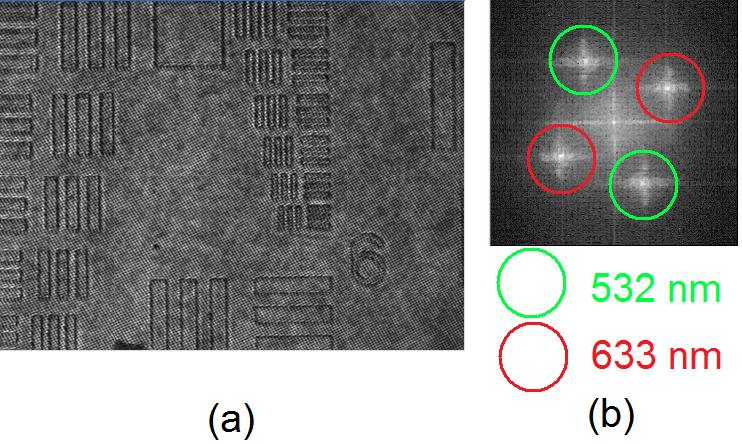
Fig. 2. Two-wavelength hologram of a USAF resolution target: (a) digital hologram (640x480 pixels) and (b) its Fourier spectrum of the hologram with the red and the green wavelengths first order components shown.

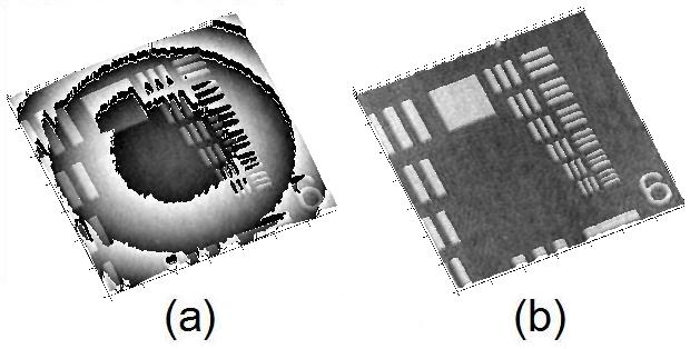
Fig. 4. The reconstructed phase image of the USAF resolution target (a) without curvature correction and (b) with curvature correction applied. The images are 174x174 μm2 (450x450 pixels).

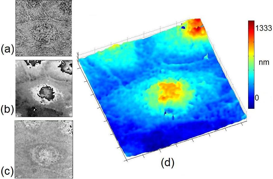
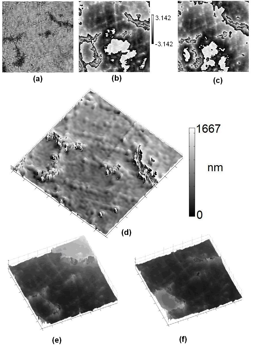
Fig. 7. Confluent SKOV-3 ovarian cancer cells: (a) amplitude image, (b) reconstructed phase for λ=532 nm, (c) dual-wavelength coarse phase image and (d) 3D rendering of fine map. All images are 92x92 μm2 (240x240 pixels).


A Khmaladze, A Restrepo-Martinez, MK Kim, R Castaneda, & A Blandon, “Simultaneous dual-wavelength reflection digital holography applied to the study of the porous coal samples,” Appl. Opt. 47, 3203-3210 (2008). > pdf

We present a simultaneous dual-wavelength phase-imaging digital holographic technique demonstrated on porous coal samples. The use of two wavelengths enables us to increase the axial range at which the unambiguous phase imaging can be performed, but also increases the noise.We employ a noise reduction“fine map” algorithm, which uses the two-wavelength phase map as a guide to correct a single-wavelength phase image. Then, the resulting noise of a fine map is reduced to the level of single-wavelength noise. A comparison to software unwrapping is also presented. A simple way of correcting a curvature mismatch between the reference and the object beams is offered.

Fig. 9. Images of a porous coal sample: (a) amplitude image; phase maps reconstructed at (b) λ1 ¼ 532nm and (c) λ2 ¼ 633nm; (d) 3D rendering of the dual-wavelength phase map; software unwrapped phase maps reconstructed at (e) λ1 ¼ 633nm and (f) λ2 ¼ 532nm for comparison. All image sizes are 98 × 98 μm2. The vertical scales of the phase maps are in radians.


D. Parshall & M.K. Kim, “Digital holographic microscopy with dual wavelength phase unwrapping”, Appl. Opt. 45, 451-459 (2006). > pdf

We apply the techniques of digital holography to obtain microscopic three-dimensional images of biological cells. The optical system is capable of microscopic holography with diffraction-limited resolution by projecting a magnified image of a microscopic hologram plane onto a CCD plane. Two-wavelength phase-imaging digital holography is applied to produce unwrapped phase images of biological cells. The method of three-wavelength phase imaging is proposed to extend the axial range and reduce the effect of phase noise. These results demonstrate the effectiveness of digital holography in high-resolution biological microscopy.


J. Gass, A. Dakoff, & M.K. Kim, ““Phase imaging without 2pi-ambiguity by multiple-wavelength digital holography”, Opt. Lett. 28, 1141-1143 (2003). > pdf

We present a phase-imaging method with an axial range that can in principle be arbitrarily large compared to the wavelength and does not involve the usual phase unwrapping by detection of phase discontinuity. The method consists of the generation and combination of two phase maps in a digital holography system by use of two separate wavelengths. For example, we reconstructed the surface of a spherical mirror with ~10-nm axial resolution and an axial range of ~3 um.

.