We demonstrate a method of optical tomography for surface and sub-surface imaging of biological tissues, based on the principle of wide field optical coherence tomography and capable of providing full-color three-dimensional views of a tissue structure. Contour or tomographic images are obtained with an interferometric imaging system using broadband light sources. The interferometric images are analyzed in the three color channels and recombined to generate 3D microscopic images of tissue structures with full natural color representation. In contrast to most existing three-dimensional microscopy methods, the presented technique allows monitoring of tissue structures close to its natural color, which may be useful in physiological and pathological applications.
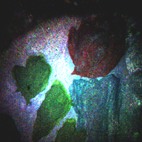 . .  . . 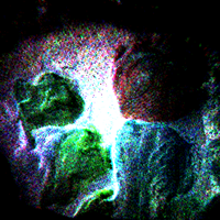
[QuickTime movies] Color WFOCT movies of a painted coin surface: (a) (1.17MB) xy-section images; (b) (0.26MB) xz-section images; (c) (0.90MB) 3D perspective views. (image volume = 7.2 mm x 7.2 mm x 335 mm; voxels = 480 x 480 x 67; voxel volume = 15 mm x 15 mm x 5 mm)
|